52+ Nerves In Neck Diagram
One pair of coccygeal Co1 nerves meets in the area of the tailbone. The spinal column contains about.

Nerve Structure Anatomy And Diagram Getbodysmart
Web The first nerve root exits between S1 and S2.
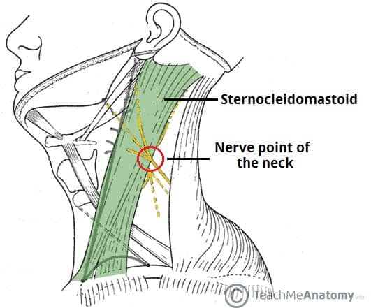
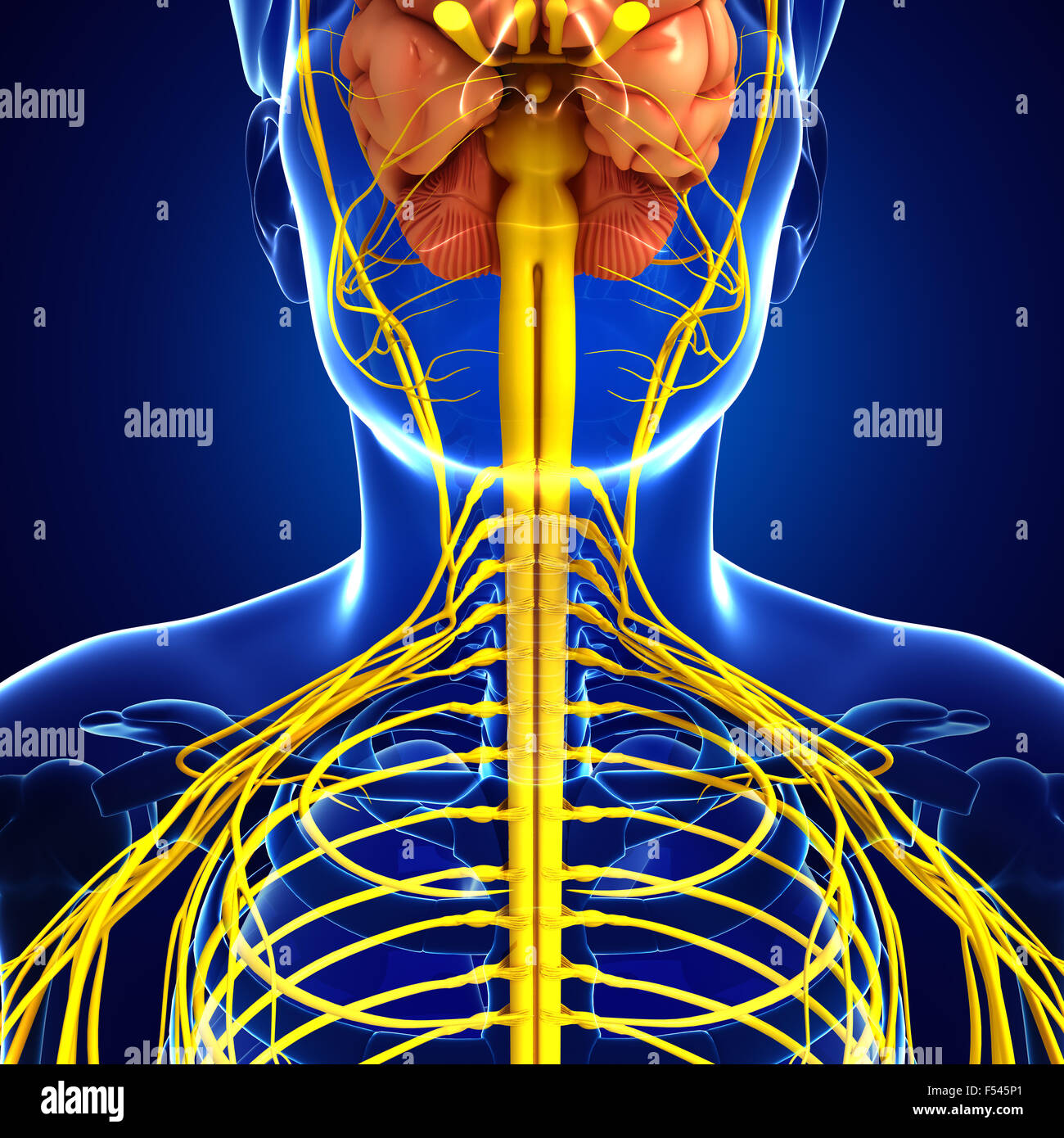
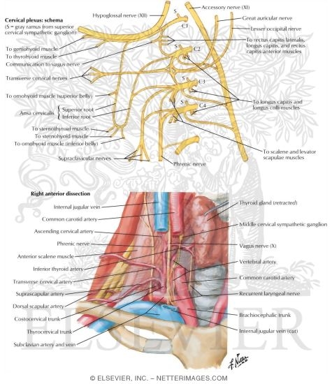
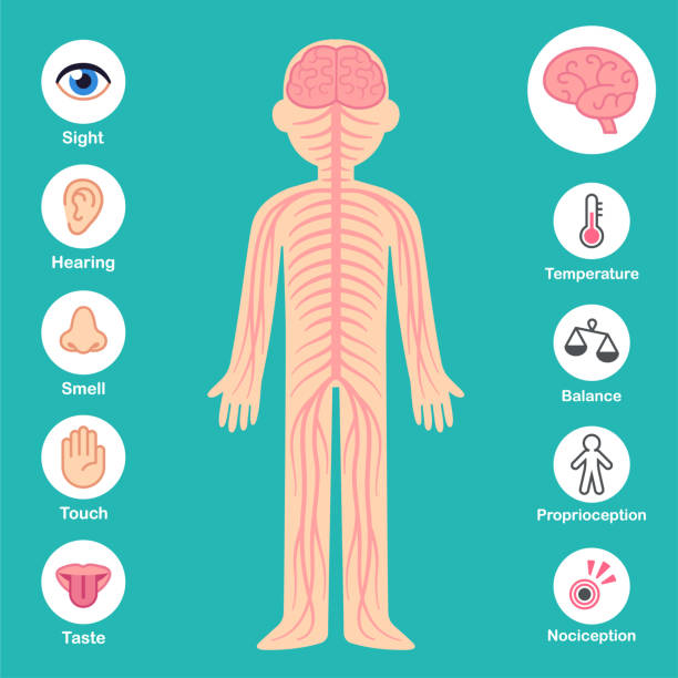
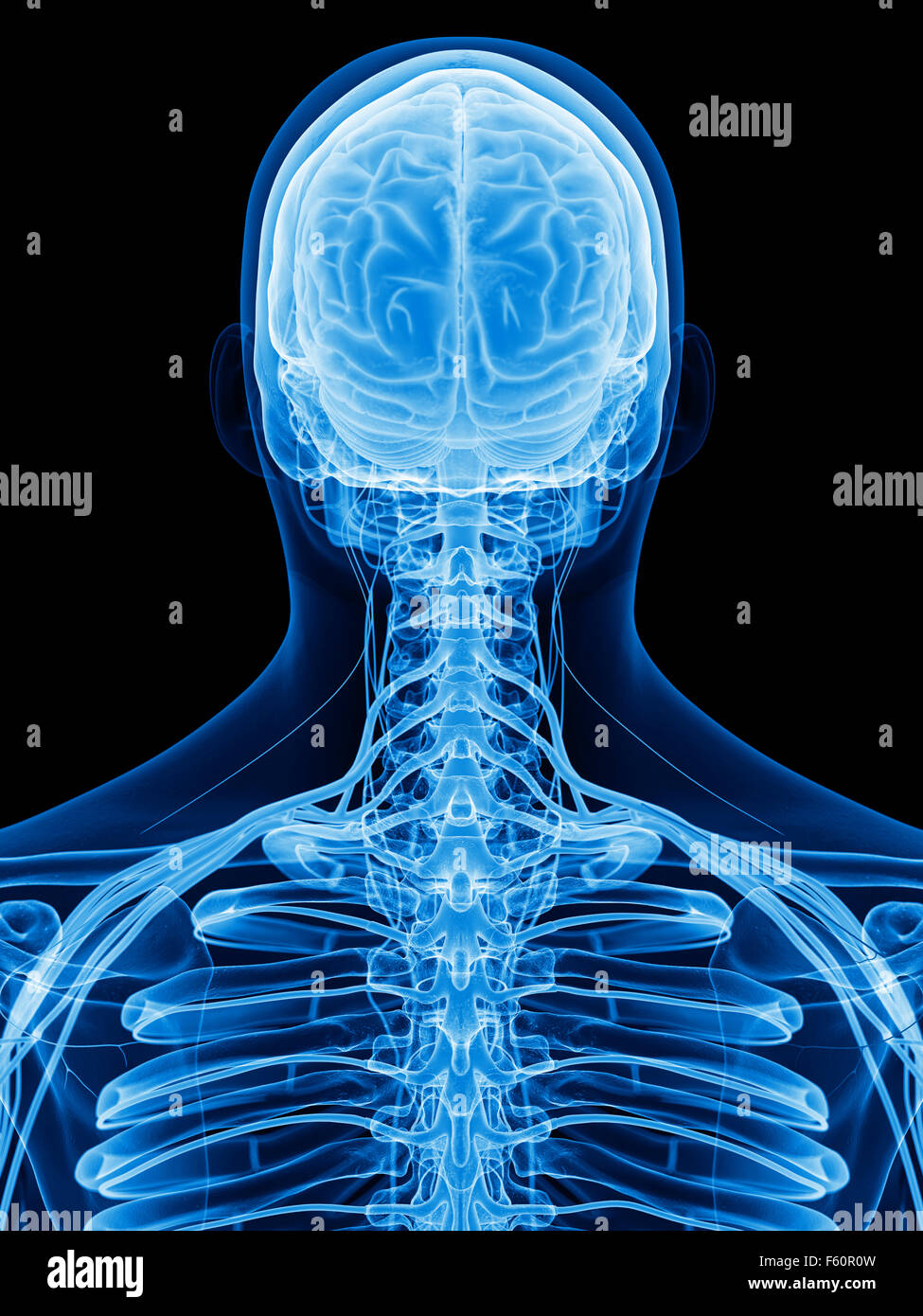
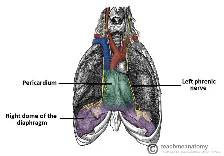
. Web This section on the nerves of the neck discusses the anatomy of the cervical plexus and the phrenic nerves. Web The spinal cord is a bundle of nerves that carry electrical signals between the brain and the rest of the body. Sweat glands of the forehead.
The middle muscle layer Iliocostalis. The outermost muscle layer Tiny muscles that connect one vertebra to. In fact there are twenty three in total some of which are.
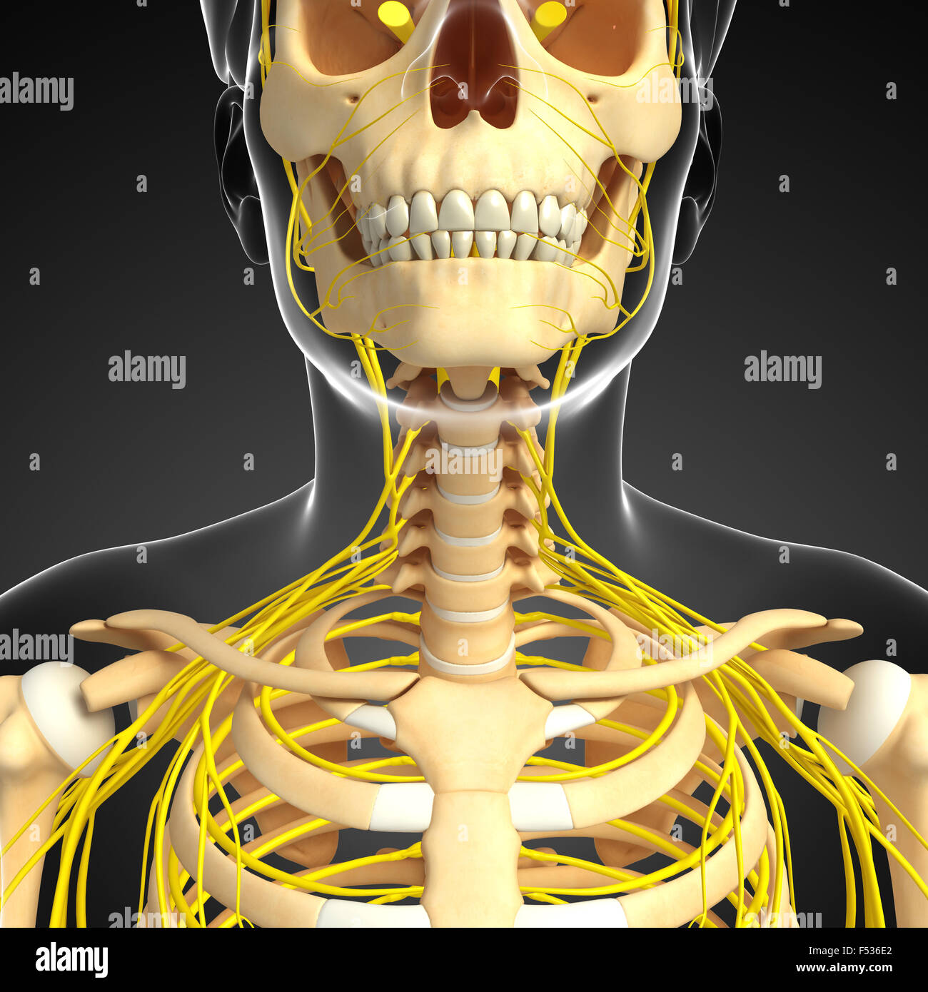
Web Neurons or nerve cell are the main structural and functional units of the nervous systemEvery neuron consists of a body soma and a number of processes. Web Some important structures contained in or passing through the neck include the seven cervical vertebrae and enclosed spinal cord the jugular veins and carotid arteries part of. By way of the peripheral nervous system PNS nerve impulses.
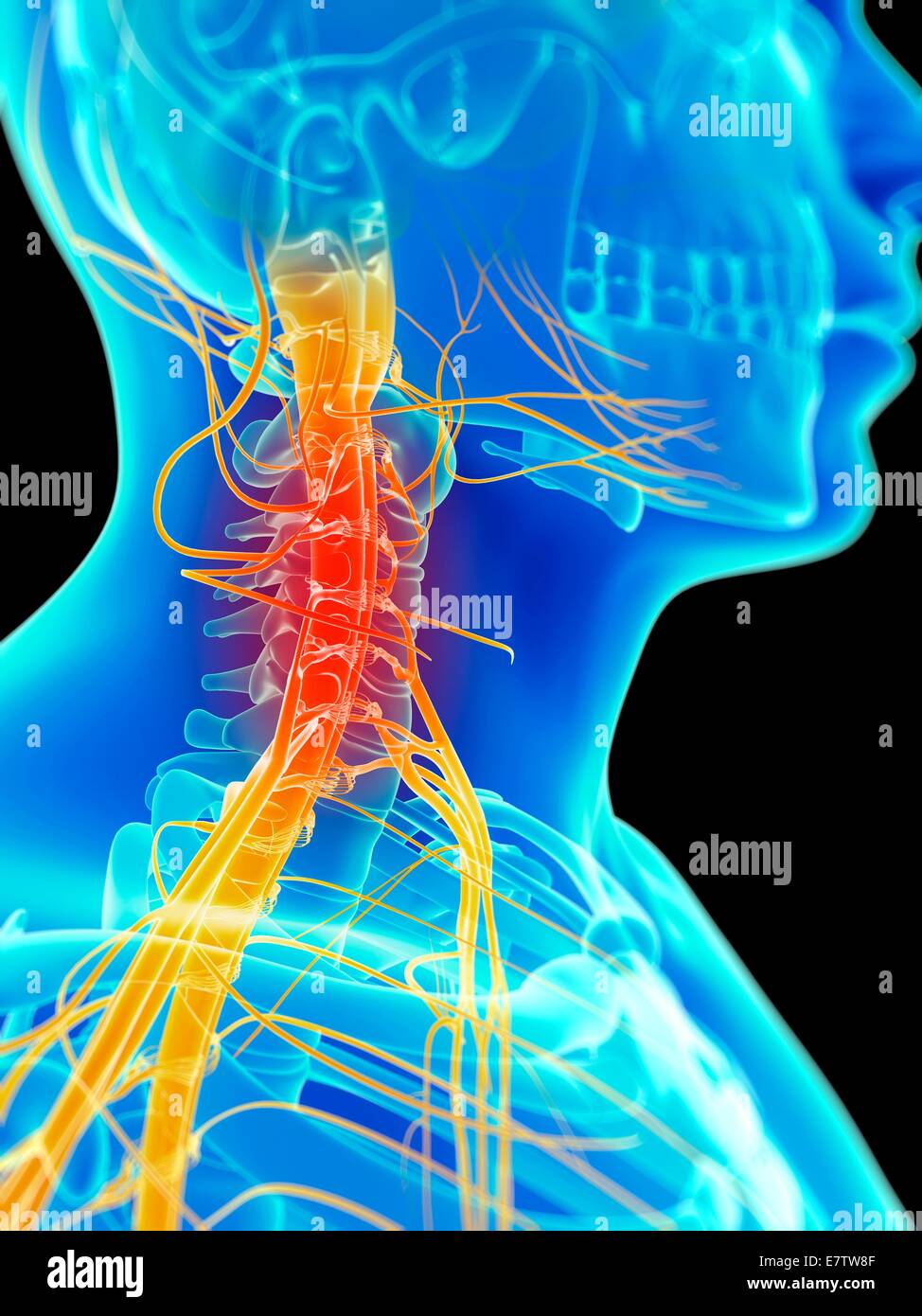
Web Main bones of the skull As you can see from the above skull diagram there are quite a lot of skull bones. Arrector pili muscles of forehead and anterior scalp. Web Cervical radiculopathy also known as pinched nerve is a condition that results in radiating pain weakness andor numbness caused by compression of any of the nerve roots in.
Web Rectus Capitis Lateralis Helps the neck to bend to the side. There are three primary occipital. The innermost muscle layer Longissimus.
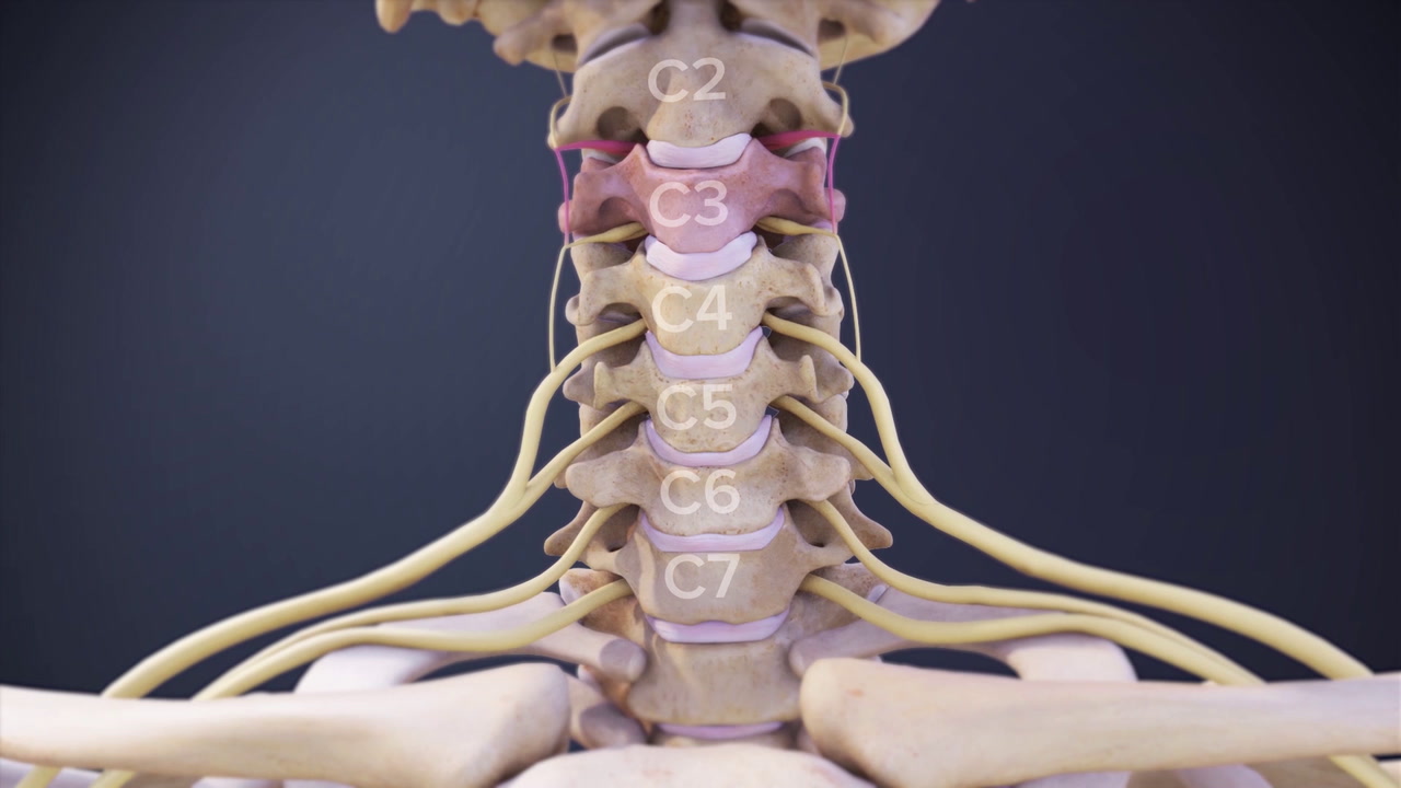
Scalene Muscles Responsible for lifting the first and second ribs ie. Web vascular smooth muscle of the brain orbit forehead upper nasal cavity. Web The occipital nerves are a group of nerves that branch from the C2 and C3 spinal nerves which help control the neck and head.
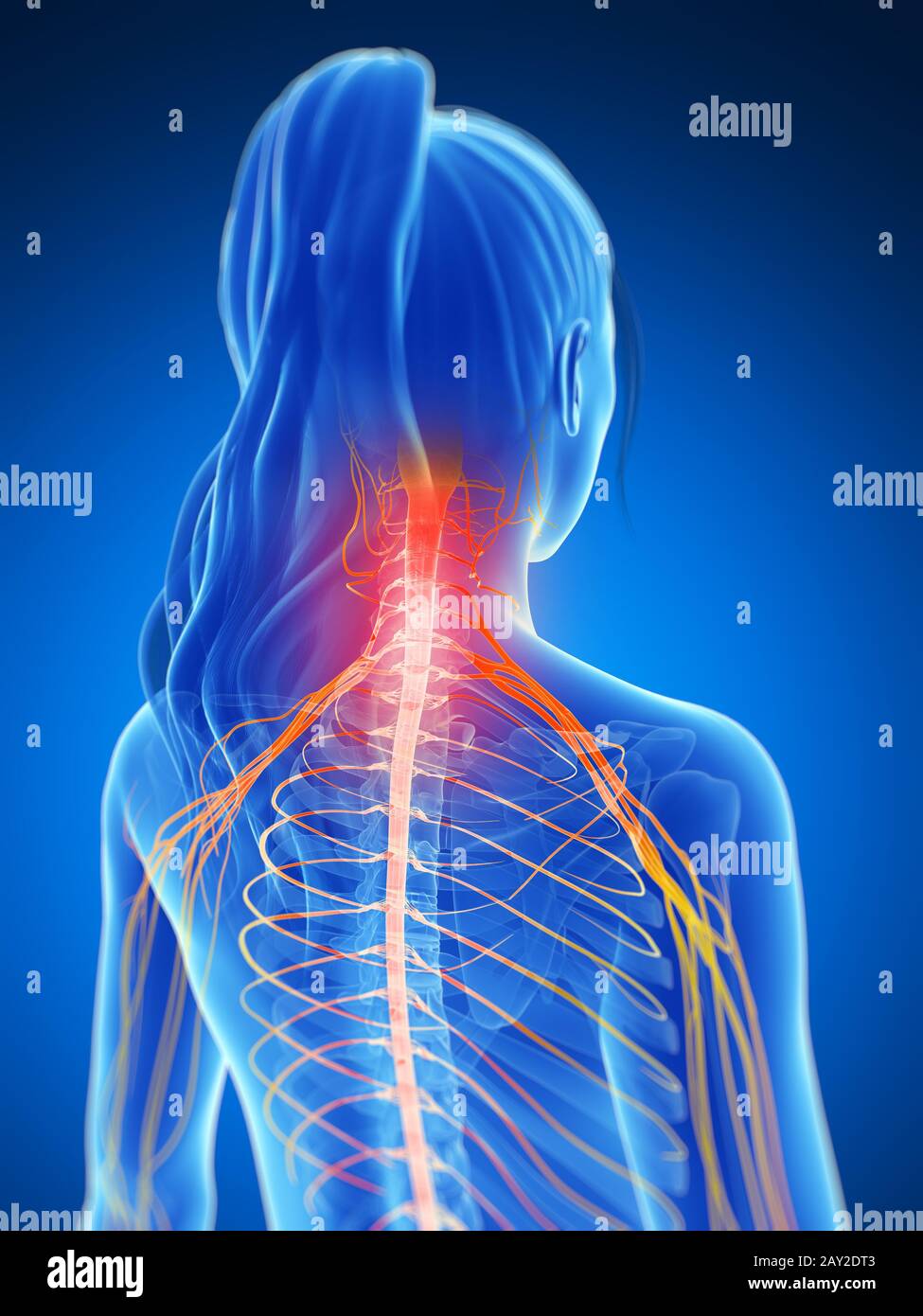
Web Neck Anatomy Area Diagram Body Maps Human body Neck Neck The neck is the start of the spinal column and spinal cord. Symptoms of a pinched nerve in the neck can include neck pain hand and arm numbness shoulder weakness. Web Spinalis.
Web Thyroid gland anatomy Hyoid bone Sources Show all Neck spaces The content of the neck is grouped into 4 neck spaces called the compartments. The cervical plexus is a network of nerves which forms from the anterior. One of the most important jobs of the cervical spine is to protect the.

Veetha Collection
![]()
Transverse Cervical Nerve Earth S Lab
Human Neck Nerves Hi Res Stock Photography And Images Alamy
Human Neck Nerves Hi Res Stock Photography And Images Alamy
Human Neck Nerves Hi Res Stock Photography And Images Alamy
The Cervical Plexus Spinal Nerves Branches Teachmeanatomy
Nerves And Vessels Of Neck

Diagram Of Superfical Nerves Of The Neck
Human Neck Nerves Hi Res Stock Photography And Images Alamy
330 Neck Nerves Stock Photos Pictures Royalty Free Images Istock
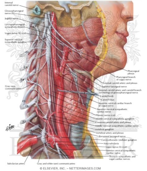
Autonomic Nerves In Neck
Human Neck Nerves Hi Res Stock Photography And Images Alamy

Nerves Of Oral Head And Neck Regions Anatomy Zygomaticotemporal Branch Of Zygomatic Nerve Cn V2 Deep Tempora Nerve Anatomy Human Anatomy Body Anatomy
Nerves Of The Neck Teachmeanatomy

Autonomic Nerves In Neck
Cervical Spinal Nerves Spine Health

7 226 Neck Nerve Images Stock Photos 3d Objects Vectors Shutterstock